
The shoulder is the most mobile joint in the body. The challenge with such extensive mobility, however, is that mechanical support and function relies heavily on the soft tissues. Consequently there are more soft-tissue disorders in this region. Bursitis and rotator cuff pathology are common diagnoses for soft-tissue shoulder pain. While both of these occur, other conditions can mimic these more common complaints. In situations like this treatment may be ineffective because they target the wrong cause. Damage or impairment to the suprascapular nerve can easily mimic a number of other shoulder complaints. Because of the location and function of this nerve, massage therapy can be a beneficial means of addressing suprascapular nerve disorders. Let’s take a look at the structure and function of this nerve, which often gets ignored in the evaluation and treatment process.
Anatomy
The suprascapular nerve is a relatively small and short nerve. Unfortunately it has to bypass several anatomical obstacles along its path and these obstacles are what make the nerve vulnerable to compression and tension injury. The suprascapular nerve originates from the C5 and C6 nerve roots and is a mixed nerve, meaning it carries both motor and sensory fibers. It provides sensory innervation to the posterior and lateral aspect of the shoulder and motor innervation to the supraspinatus and infraspinatus muscles.
The main portion of the nerve begins where it branches off of the superior trunk of the brachial plexus in the cervical region (Image 1). It branches off of the brachial plexus relatively high up along the bundle of nerves so it is less susceptible to a number of the other compression pathologies that affect lower portions of the brachial plexus.
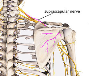
Image 1: Suprascapular nerve and its origin from brachial plexus
After branching off of the superior trunk of the brachial plexus it travels across the top of the shoulder on its way to the superior and lateral aspect of the scapula. The first primary anatomical obstacle it encounters is the narrow passage through the suprascapular notch (Image 2). The edges of the scapula along the suprascapular notch can be somewhat sharp and they may cause damage to the nerve when it is pulled taut against the edge of the notch.
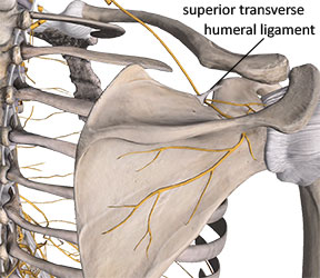
Image 2 Suprascapular notch with superior transverse ligament
There is a small ligament that spans the opening at the top of the suprascapular notch. This ligament is called the transverse scapular ligament. Because it spans the opening of the suprascapular notch it essentially creates a small foramen that the nerve must travel through (Image 2).
The transverse scapular ligament can sometimes become ossified, creating an even more rigid border for this narrow channel that the nerve passes through.1. There is also some indication that the suprascapular notch may narrow with aging. 2 Ligament ossification and narrowing of the opening at the suprascapular notch are both contributing factors to suprascapular nerve compression.
After the nerve courses through the suprascapular notch, branches extend out that innervate the supraspinatus muscle. Some of the remaining branches that pass the spine of the scapula and go down to the infraspinatus region may actually pass through the supraspinatus muscle; this is another potential site of nerve entrapment.
After passing the supraspinatus region, the nerve makes a sharp bend as it curves around the spinal glenoid notch (Image 3). Where the nerve bends around the rigid bony border of the spinoglenoid notch, it is vulnerable to compression.
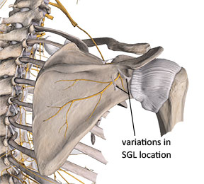
Image 3 Spinoglenoid notch and spinoglenoid ligament (SGL)
In up to 50% of people there is a connective tissue band called the spinoglenoid ligament that forms a second narrow tunnel where the nerve must pass (Image 3). 3 This ligament is also designated in some resources as the inferior transverse scapular ligament. Some anatomical references show this small ligament covering the suprascapular nerve farther along its path away from the spinoglenoid notch (Image 3). Because these small ligaments do not appear in all individuals, it is possible that there may be multiple locations where these anatomical anomalies occur. Regardless of their location, the impact on compressing the suprascapular nerve is similar in both situations.
Pathophysiology and Etiology
The suprascapular nerve has very little room for additional movement as it passes by these anatomical obstacles. Nerves must be somewhat mobile and able to slide relative to adjacent structures. Some scapular movements put significant tensile (pulling) stress on the suprascapular nerve and may lead to nerve pathology.
Protraction and abduction both pull the nerve against rigid bony borders and may cause neural tension or compression. Elevation and rotation of the arm can also put traction forces on the nerve because of the movement of adjacent muscles that pull on nerve fibers. A number of shoulder pathologies, such as fractures of the clavicle or scapula, soft-tissue cysts or masses, or soft-tissue injuries like rotator cuff tears, can also lead to suprascapular nerve compression. Sometimes these shoulder injuries distort the pathway of the nerve, causing neural compression or tension.
Frequent overhead work or any positions that involve repeated abduction and external rotation put the nerve in a vulnerable position. Suprascapular neuropathy is common in many sporting activities. It is reported with frequency in volleyball players because of the vigorous overhead motions with the dominant hand during volleyball play.4.
Suprascapular neuropathy can also cause other shoulder problems from biomechanical dysfunction. Nerve compression may cause atrophy of supraspinatus and infraspinatus muscles, which then causes dysfunctional mechanics of the rotator cuff. For example, the infraspinatus and teres minor muscles play a major role in decelerating momentum of the arm at the end of a throwing motion. If the infraspinatus is weak due to nerve compression, it can’t generate the same eccentric force to decelerate the arm. Weakness of the posterior rotator cuff complex can lead to tendinosis in the affected tendons. When these muscles are weak, more of that deceleration forces transfer to the joint capsule tissues and that may stress these capsular tissues.
Muscular atrophy and weakness is a primary symptom from suprascapular neuropathy. However, motor symptoms do not occur in isolation because there are also sensory fibers within the suprascapular nerve. Impaired sensory signals that may occur from suprascapular neuropathy include increased shoulder pain in the posterior or lateral shoulder region. There may also be tingling, numbness, or other altered sensory sensations in the region.
Identifying nerve compression
It is challenging to identify suprascapular neuropathy because its symptoms are similar to many other shoulder complaints. For example, people may report lateral shoulder pain along with difficulty bringing the shoulder into abduction. It is common for this pattern to be diagnosed as rotator cuff pathology or subacromial bursitis without a thorough exploration of other possible causes. These same symptoms could be attributed to suprascapular neuropathy. Difficulty abducting the arm occurs because the supraspinatus muscle, which is essential for abduction, is not getting adequate motor signals. Posterior and lateral shoulder pain occurs as the nerve gets further stretched during the motion of abduction.
Key factors to look for in the client history include long periods of holding the shoulder in abduction or significant repetitive motion where the shoulder is brought into abduction or protraction numerous times. Pain, usually described as sharp, will generally accompany that position.
Observation of the posterior shoulder region may give helpful clues in more advanced stages of the condition. Infraspinatus atrophy that occurs from suprascapular nerve compression is often invisible because there are few other muscles overlying it. Infraspinatus atrophy may appear as a slightly hollowed out depression on the posterior aspect of the shoulder right over the infraspinous fossa.
Impaired motor signals to the affected muscles from suprascapular nerve compression can also be evaluated with manual resistive tests. Perform the test on the healthy side first to establish a normal baseline. Resisted abduction will test function of the supraspinatus while resisted lateral rotation will test the integrity of the infraspinatus. If the resisted motion is weak there may be impairment of the suprascapular nerve. There are other muscles which assist these two rotator cuff muscles in each of their actions, so suprascapular nerve compression will not completely stop active motion, it will simply appear to be weaker than the healthy side.
While the report of pain from suprascapular nerve compression is usually in the posterior and lateral shoulder region, it can also sometimes be felt down the upper extremity, especially along the area innervated by the radial nerve or its branches. It follows patterns of the radial nerve because the suprascapular and radial nerves share a similar pathway of origin at the C5 and C6 nerve roots and the superior trunk of the brachial plexus.
Nerve compression or tension problems can also be further identified by increased sensitivity to palpation along the nerve’s path. If pressing into the distal region of the supraspinatus muscle or the most lateral aspect of the infraspinatus near its musculotendinous junction reproduces this pain, there is a likelihood of suprascapular nerve involvement. Interestingly, common trigger point maps show irritable trigger points just below the spine of the scapula that tend to refer pain or other sensations down the upper arm along the same path of the radial nerve (Image 4). Palpating the posterior shoulder area where these active trigger points exist could likely compress the suprascapular nerve and that could be a partial explanation for this pain pattern that appears down the upper arm.
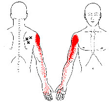
Image 4:
Common trigger points in the infraspinatus
Treatment
Suprascapular neuropathy can be a challenging problem to address because there are few things that can be done for it. The most important factor is to identify any biomechanical patterns, such as keeping the arms in abduction for long periods or excessive and repetitive motion that may be aggravating the nerve compression. Limiting those aggravating factors is the first strategy for addressing any nerve compression problem.
As noted earlier, branches of the suprascapular nerve may course directly through the supraspinatus muscle and may be bound or restricted by the other tissues that interface with the nerve. Consequently it is helpful to reduce any hypertonicity in the supraspinatus or infraspinatus muscles to encourage full freedom and mobility of the nerve so it can easily glide in relation to adjacent structures.
One of the most effective ways to help reduce hypertonicity in the supraspinatus muscle is through deep stripping techniques applied directly to the supraspinatus in a longitudinal direction along its length. It is difficult to access the most distal portion of the supraspinatus near its musculotendinous junction as the muscle goes under the acromion process at the acromioclavicular joint. However the area of potential suprascapular nerve restriction is more proximal to the region and is accessible through massage treatment.
When performing this stripping technique make sure the contact (finger, thumb, pressure tool) remains anterior to the large bundle of the upper trapezius. If you simply perform the stripping technique across the top of the shoulder without getting anterior to the trapezius you will simply be working on the trapezius and will give very little pressure down into the supraspinatus.
Once the contact point is established anterior to the trapezius, perform a slow longitudinal stripping technique along the length of the supraspinatus (Image 5). This can be performed moving medial to lateral or lateral to medial. One of the key goals of this longitudinal stripping technique is to help encourage mobility between the supraspinatus and the affected nerve. You can further enhance mobility by either actively or passively moving the client’s arm through abduction and adduction as you perform the stripping technique. Moving the muscle as you work on it helps increase greater tissue flexibility as well as mobility of all surrounding structures.
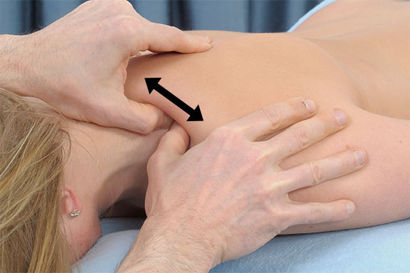
Image 5
Deep stripping to the supraspinatus
A similar technique can be applied to the infraspinatus to reduce hypertonicity and increase mobility between the suprascapular nerve and the infraspinatus muscle. With the client in a prone position, perform a slow stripping technique along the length of the infraspinatus muscle in a fan-shaped pattern with a small contact surface such as a finger, thumb, or pressure tool (Image 6). Make sure to check in with the client frequently about the appropriate depth of pressure and be sure not to apply so much pressure that any of the existing neurological sensations are increased. Be particularly cautious when applying pressure just inferior to the spine of the scapula, as that is where the primary area of nerve entrapment is usually located.
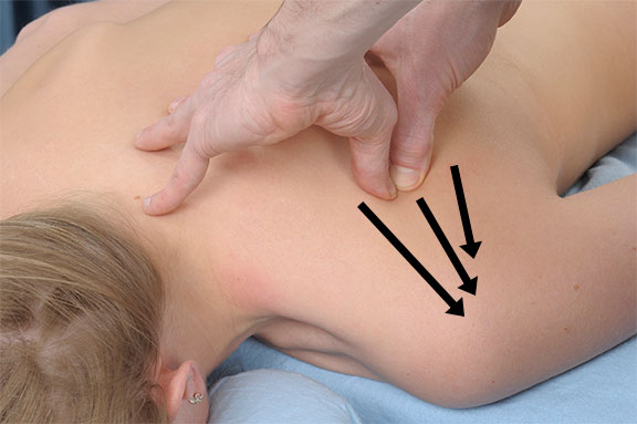
Image 6:
Deep stripping to the infraspinatus
Just like with the supraspinatus, it may be helpful to conduct active or passive movement during treatment to encourage full freedom of movement between the infraspinatus and suprascapular nerve. Often it is most effective to have the client moving the arm in medial rotation, so that the muscle is lengthening as the technique is applied. Make sure the client is close to the edge of the table to allow full freedom of movement through the entire range of motion in medial and lateral rotation as you apply this technique (Image 7).
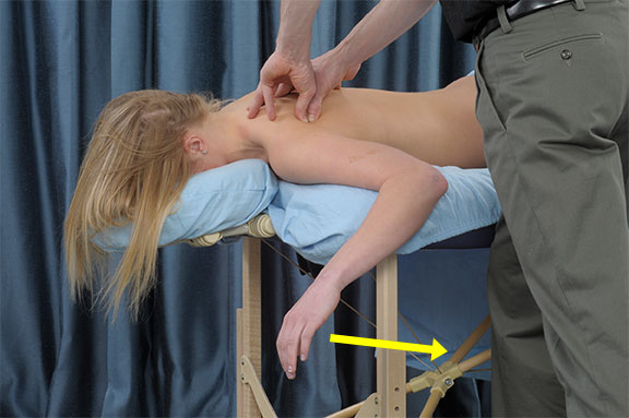
Image 7:
Deep stripping to the infraspinatus during active medial rotation
While suprascapular neuropathy is not as common as other nerve compression problems like carpal tunnel syndrome, it is still valuable to know about its existence and recognize some of the key factors. It is particularly beneficial for massage therapists to be aware of this problem because there are few other treatment approaches that are as effective as massage in mobilizing the nearby tissues and helping the nerve’s return to optimum function. The massage therapist who is able to skillfully apply evaluation and treatment strategies for addressing this problem will be a valuable player in your client’s return to full function and health – and that means much more success for your practice!
References
- Pecina M, Markiewitz A, Krmpotic-Nemanic J. Tunnel Syndromes: Peripheral Nerve Compression Syndromes. Boca Raton: CRC Press; 2001.
- Yamakado K. The suprascapular notch narrows with aging: a preliminary solution of the old conjecture based on a 3D-CT evaluation. Surg Radiol Anat. 2016;38(6):693-697. doi:10.1007/s00276-015-1614-5.
- Kharay S, Sharma A, Singh P. Unusual morphology of scapulae: incidence and dimensions of ossified ligaments and supraspinous bony tunnels for clinical consideration. Singapore Med J. 2016;57(1):29-32. doi:10.11622/smedj.2015103.
- Witvrouw E, Cools A, Lysens R, et al. Suprascapular neuropathy in volleyball players. Br J Sports Med. 2000;34(3):174-180. http://www.ncbi.nlm.nih.gov/pubmed/10854016.