
Updated 01/31/2024
Introduction
Back pain contributes significantly to disability and frequently leads people to seek out massage therapy. Often, back pain is diagnosed as “non-specific” due to the absence of identifiable tissue pathology. However, noted back pain researcher Stuart McGill, PhD, has often advocated that one of the main reasons there are so many diagnoses of “non-specific back pain” is that people didn’t take enough time to be thorough in their assessment. The story of “back mice” may be one of those situations.
Physiology of Back Mice
So, what are “back mice?” They are moveable fibro-fatty nodules found primarily near the sacroiliac region at the top of the iliac crest. They are palpable as rubbery nodules beneath the skin and are also called episacroiliac lipomas. The precise reason for their prevalence in this area remains unclear. Research involving cadavers suggests their formation is linked to fatty tissue herniating through fascial layers, potentially related to the complex fascial structures in the region.
The primary cause of the soft-tissue nodules found in the sacroiliac region is not entirely understood. Studies on cadavers indicate their occurrence in areas where fatty tissue protrudes through fascial layers, possibly linked to the multiple fascial layers in this area. A case study in a recent paper highlights this, featuring a 47-year-old woman with bilateral low back pain, unattributable to any significant back tissue dysfunction. Her symptoms also included intermittent numbness and cramping in the posterior thigh.(1)
A comprehensive physical examination revealed no major neurological issues, nerve root pathology, or spinal structure involvement. However, there were palpable soft-tissue nodules in the sacroiliac region. Pressure on these nodules reproduced the patient’s primary pain. The successful treatment involved corticosteroid and anesthetic injections into the nodules, effectively alleviating her pain.
Back mice, often encountered by massage therapists, are physiologically characterized as fatty herniations through fascial layers. In the case study mentioned above, the primary treatment was corticosteroid and anesthetic injections. Interestingly, these injections did not seem to alter the fatty nodules physically; instead, they targeted the alleviation of pain. The patient experienced complete symptom resolution following this treatment.
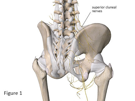
The successful relief of symptoms through a pain management approach, without physically modifying the nodules, suggests that the pain associated with back mice might be primarily neurological. It raises the question of whether the nodules exert pressure on nearby nerves, such as the cluneal nerves (Figure 1), or other local cutaneous nerves, causing the pain.
Orthopedic Medical Massage Treatment
Massage therapists, with their advanced palpation skills, often identify fibro-fatty nodules in clients who report back pain. An important practice is to palpate these nodules to see if they exacerbate the specific back pain reported. If they do, these nodules might be partially or completely causing the pain.
This raises a key question: what role can massage play in alleviating discomfort from these ‘back mice’? Deep pressure on the nodules may not be effective. However, if the nodules are irritating superficial nerve structures like the cluneal nerves, light massage techniques aimed at mobilizing superficial fascial tissues and reducing nerve irritation could be beneficial.
When encountering clients with back, posterior pelvis, or posterior thigh pain who also have these episacral lipomas, consider these ‘back mice’ as a potential pain source. The challenge remains in devising an effective treatment strategy for these cases. Now if we could only build a better mouse trap…
- Bicket MC, Simmons C, Zheng Y. The Best-Laid Plans of “Back Mice” and Men: A Case Report and Literature Review of Episacroiliac Lipoma. Pain Physician. 2016;19(3):181-188. http://www.ncbi.nlm.nih.gov/pubmed/27008292.